
European Society of Toxicologic Pathology
ESTP Congress 2003: Case No 2
case index | << previous case | next case >>
|
|


Abstract
C.I. Rühl-Fehlert1, X. Palazzi2, M. Dorgerloh3, and C. Bomke4
1 Bayer HealthCare AG, Department of Toxicologic Pathology, Wuppertal, Germany
2 Bayer CropScience AG, Department of Pathology, Sophia Antipolis, France
3 Bayer CropScience AG, Institute for Ecotoxicology, Monheim, Germany
4 Dr. Knoell Consult, Mannheim, Germany
Key words: Fish, ovary, microsporidia, pleistophora
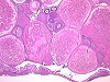
Histopathological investigation of the ovaries of a clinically healthy adult female fathead minnowa showed microsporidian infestation in the oocytes. Spores were observed within the yolk of mature oocytes. The affected oocytes often showed signs of degeneration with infolding or disintegration of the oocyte membrane. In the interstitium, a granulomatous inflammation developed predominantly in the surroundings of infected oocytes. After complete degeneration of oocytes, spore masses were also detected within the inflammatory cells.
The animal was derived from a batch of heavily infected fathead minnows with about 70% of the females showing spores in the oocytes. In males, only small cysts were found in the deferent ducts and marginal inflammatory changes in the testes. Based on morphology and organ-specific distribution, an infection with Pleistophora spp. was assumed. The species could not be clarified by light microscopic investigations and taking into consideration varying classifications in literature. The parasite is known to affect various fish species and to cause economic losses in commercial fish farms due to lowering of reproductive rates since the infection eventually leads to severe ovarian destruction. The mode of infestation is yet unclear, horizontal, and vertical transmission via infected oocytes as well as autoinfestation have to be taken into account.