 |
 | |
| Species: |
Dog |
| Strain/breeder: |
Beagle |
| Sex: |
Male |
| Age: |
1 year |
| Study type: |
1 month oral |
| Treatment: |
Control (tap water) |
| Animal status: |
No clinical abnormality |
| Clinical findings: |
ECG showed slight prolongation of the PR interval on day 22. No remarkable findings on hematology, clin. chemistry or urinanalysis. |
| Organ(s): |
Intestine (jejunum, colon, rectum) (1a), salivary glands, axillary lymph node (1b), thyroids (1c), Kidneys (1d), skin (1d). |
Gross
finding(s): |
Nothing abnormal detected |
| Staining: |
H&E |
| Literature: |
 |
Jacobs et al. (2002). In: Tumors in Domestic Animals, Meuten DJ (editor), Iowa State Press, pp 170–172 |
|
|
|
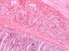
Fig. 1 (96k)
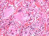
Fig. 2 (99k)
|
|
Abstract
Granulomatous inflammation of unknown etiology in multiple organs in a dog
S. Bjurström
AstraZeneca R&D Söderälje, Safety Assessment, Södertälje, Sweden
Key words: Granulomatous inflammation, histiocytosis, dog
This case report describes a spontaneous granulomatous inflammatory reaction in multiple organs a 1-year-old male beagle dog from a control group (treated with tap water) in a 1-month oral toxicity study. The dog showed no clinical sign of disease. ECG showed slight prolongation of the PR interval. There were no remarkable findings on hematology, clinical chemistry or urine analysis. At necropsy, no remarkable gross findings were observed. The histological examination revealed multifocal granulomatous inflammation in numerous organs and tissues. Heart, kidneys, stomach, intestines, pancreas, salivary glands, thyroids, gall bladder and skin (sebaceous glands) were among the more heavily affected organs while other organs like lungs, skeletal muscles and brain tissue appeared not to be involved. The inflammation was characterized by multifocal cell-rich infiltrates of mononuclear inflammatory cells (lymphocytes, plasma cells, histiocytes), often showing a granulomatous appearance with admixture of large, multinucleated macrophages. Staining with Ziehl-Nielsen for tubercle bacilli was negative. A probable diagnosis is histiocytic proliferative disease ( i.e. systemic histiocytosis) although the present case does not fulfill all diagnostic criteria (Jacobs et al.: in Tumors in Domestic Animals, D. J. Meuten (ed), Iowa State Press, 2002; pp. 170-172).
|
case index | << previous case | next case >>
|
|