
European Society of Toxicologic Pathology
ESTP/GTP meeting 2002: Case No 3
case index | << previous case | next case >>
|
|

Abstract
A. ROMEIKE, L. LONGEART, J-L. LENET, M. ANDREU and M.-T. MASSON
Pfizer Global R & D, Amboise Laboratories, BP 109, 37401 Amboise Cedex, France
Key words: skin, mycosis fungoides, epidermotropic lymphoma, rat, Pautrier's microabscesses, T8 lymphocytes, SD rat
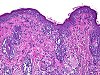
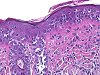
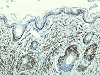
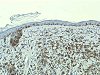
Seven spontaneous cases of cutaneous epidermotropic lymphomas (mycosis fungoides) occurred in 2 male and 5 female SD rats that belonged to control or treated groups from 3 different 2-year carcinogenicity studies. Histologically, the lesions consisted of a loose lichenoid and periadnexal dermal infiltrate of neoplastic cells. The infiltrate was composed of round mononuclear cells of about 12-15 µm in diameter, with scant cytoplasm and indented nuclei. The presence of neoplastic cells within the epidermis and/or hair follicle epithelium, generally grouped in clusters (Pautrier's microabscesses), was a constant feature. Mitoses were rare. Electron microscopically the neoplastic cells were typical lymphocytes: indented nucleus with marginated chromatin and an occasional nucleolus, cytoplasmic projections, and a few organelles (mainly mitochondria, reticulum profiles, and ribosomes). The results of the immunhistochemical labelling with pan-T (OX34), non-B (W3/13), and CD8 (OX8) markers suggested that these lymphomas were of T8 origin. Among laboratory animals, epidermotropic cutaneous lymphomas have been described in Syrian hamsters, mice and SD rats.