 |
 | |
| Species: |
Dog |
| Strain/breeder: |
Mixed breed German sheperd dog |
| Sex: |
Male |
| Age: |
9 years |
| Study type: |
- |
| Treatment: |
- |
| Animal status: |
Humanely destroyed |
| Clinical findings: |
Sudden blindness, pyrexia, hyperaemia of oral mucosa |
| Organ(s): |
Oral mucosa, Brain |
Macroscopic
finding(s): |
Oral mucosa: no abnormalities detectable
Brain: Tumor mass in the ventral midline of the diencephalon effacing the optic chiasm and invading the hypothalamic area |
| Staining: |
H&E, paraffin wax-embedded |
|
|

Fig. 1 (90k)
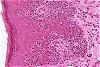
Fig. 2 (80k)
|
|
Abstract
Cutaneous epitheliotropic lymphosarcoma in a dog with central nervous system metastasis
S. CZASCH1, K. RISSE2 and W. BAUMGÄRTNER3
1Merck KGaA, Institut für Toxikologie, Frankfurter Straße 250, 64293 Darmstadt, Germany
2Staatliches Medizinal-, Lebensmittel- und Veterinäruntersuchungsamt Mittelhessen, Gießen, Germany
3Institut für Pathologie, Tierärztliche Hochschule Hannover, Hannover, Germany
Key words: epitheliotropic T-cell lymphosarcoma, dog, CNS metastasis
This report describes an uncommon case of a cutaneous epitheliotropic T-cell lymphosarcoma with central nervous system (CNS) manifestations in a 9-year-old mixed breed German shepherd dog. The animal had a history of sudden blindness, pyrexia and multifocal areas of hyperaemia in the oral mucosa. A biopsy from the muco-cutaneous junction of the lips led to the diagnosis of an epitheliotropic lymphosarcoma and the animal was killed. At necropsy, hyperaemia in the oral mucosa was no longer detectable. In the brain, a mass effacing the optic chiasm and invading the hypothalamic area was found; histological examination revealed lymphoid tumour cell infiltration. In the epithelium of the oral mucosa, intra-epithelial lymphoid tumour cells, sometimes arranged in small clusters (Pautrier's microabscesses), in combination with a mild inflammation in the superficial dermis were observed. Oral mucosa and brain tumour cells expressed CD3 antigen, indicating their T-cell origin. This is, to our knowledge, the first report of a cutaneous epitheliotropic lymphosarcoma with CNS metastasis in a dog (CZASCH et al. J Comp Pathol 2000; 123: 59-63)
|
case index | << previous case | next case >>
|
|