 |
|
 | |
| Species: |
Monkey |
| Strain/breeder: |
Pygmy marmoset (Cebuella pygmea) |
| Sex: |
Male |
| Age: |
4 years |
| Study type: |
Case report |
| Treatment: |
- |
| Animal status: |
Died |
| Clinical findings: |
- |
| Organ: |
Liver |
Macroscopic
finding(s): |
Discolorated (uniformly tan) and enlarged |
| Staining: |
H&E |
| Literature: |
 |
Bennett BT, Abee CR, Henrickson R (1998) Nonhuman Primates in Biomedical Research. Disease, Chapters 1 and 10. Academic Press, San Diego, New York, Boston, pp 1-57 and 377-414 |
 |
Montali RJ, Scanga CA, Pernikoff D, Wessner DR, Ward R, Holmes KV (1993) A common-source outbreak of callitrichid hepatitis in captive tamarins and marmosets. J Infect Dis 167: 946-950 |
|
Montali RJ, Ramsay EC, Stephensen CB, Worley M, Davis JA, Holmes KV (1989) A new transmissible viral hepatitis of marmosets and tamarins. J Infect Dis 160: 759-765 |
|
Montali RJ (1993) In: Jones TC, Mohr U, Hunt RD (eds) Nonhuman Primates II. Springer Verlag, Berlin, Heidelberg, New York, pp 61-62 |
|
|
|

Fig. 1 (55k)
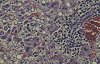
Fig. 2 (70k)
|
|
Abstract
CALLITRICHID HEPATITIS IN A PYGMY MARMOSET (CEBUELLA PYGMEA)
Aim of the study
The study describes an unusual case of hepatitis which occurred in a pygmy marmoset.
Materials & methods
Necropsy was performed on an animal which was submitted by a zoo after 12 animals died for unknown reasons. Representative tissues were formalin-fixed, routinely embedded in paraffin and stained with H&E. Several organs were freshly frozen for virus culturing and PCR methods.
Results
At necropsy, the animal showed ikteric discoloration and hepatosplenomegaly. The liver was uniformly tan and fragile. Lymph nodes were generally enlarged and hyperaemic. Histology revealed severe multifocal necrosis of liver parenchyma with periportal infiltrates of mononuclear cells and fatty degeneration. Furthermore randomly distributed acidophilic bodies could be found. Lymph nodes and spleen were depleted and necrotic. From frozen liver and spleen tissue lymphocytic choriomeningitis virus (LCMV), an arenavirus, could be isolated and confirmed by immunofluorescence and molecular techniques (PCR, sequencing).
Conclusions
This report describes a case of callitrichid hepatitis in a German zoo. The diagnosis was made on the basis of histological and virological investigations including isolation of the causative agent and comparison to other known LCMV strains. Callitrichid hepatitis is a rare but rapidly progressive viral hepatitis occurring in zoos. Outbreaks have been described in several tamarin and marmoset species. The source of infection are infected wild or neonatal laboratory mice ("pinkies"). Hemorrhagic fevers like Lassa fever in humans may be caused by similar arenaviruses and LCMV may induce disease in humans, for example after having contact with infected marmosets. This zoonotic potential has to be borne in mind.
case index | << previous case | next case >>
|
|