 |
|
 | |
| Species: |
Squirrel monkey |
| Strain/breeder: |
Saimiri sciureus |
| Sex: |
Male |
| Age: |
15 years |
| Study type: |
Natural infection |
| Treatment: |
- |
| Animal status: |
Spontaneous death |
| Clinical findings: |
Apathia |
| Organ: |
Liver, small intestine |
Macroscopic
finding(s): |
Liver: hyperemia; small intestine: pin-point mucosal hemorrhages |
| Staining: |
H&E |
| Literature: |
 |
Dubey JP, Lindsay DS (1988) Toxoplasmosis of animals and man. Boca Raton, Florida, CRC Press |
 |
Cunningham AA, Buxton D, Thomson KM (1992) An epidemic of toxoplasmosis in a captive colony of squirrel monkeys (Saimiri sciureus). J Comp Pathol 107: 207-219 |
|
Brack M, Wohlsein P, Minnemann D, Brandt HP (1998) Toxoplasmosis outbreak in ring-tailed lemurs (Lemur catta) and squirrel monkeys (Saimiri sciureus). Primate Report 50: 71-82 |
|
Wohlsein P, Brandt HP, Brack M, Peters M, Schares G, Böer M (1999) Immunhistologische und molekularbiologische Untersuchungen von nichthumanen Primaten mit disseminierter Toxoplasmose. Verh Ber Erkrg Zootiere 39: 209-214 |
|
|
|
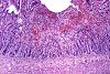
Fig. 1 (113k)
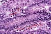
Fig. 2 (86k)
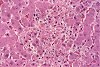
Fig. 3 (84k)
|
|
|
Abstract
EPIDEMIC DISSEMINATED TOXOPLASMOSIS IN NON HUMAN PRIMATES
Aim of the study
In a northern German wildlife park spontaneous deaths occurred in colonies of ring-tailed lemurs (Lemur catta), squirrel monkeys (Saimiri sciureus) and liszt monkeys (Sanguinus oedipus) with a generally short clinical period of apathia, anorexia and weakness. Abortions and stillbirths were observed in pregnant squirrel monkeys.
Materials & methods
To identify the cause of deaths, six lemurs, three squirrel monkeys, and three liszt monkeys were submitted for necropsy.
Results
Grossly, in some animals a slight icterus and mild serous effusion in the thoracic cavity were present. The lungs were poorly retracted and exhibited variable degrees of alveolar oedema and emphysema. The myocardium appeared mottled, the spleen was enlarged and occasional pin-point greyish foci were detected in the liver. In some animals poorly demarcated ecchymal hemorrhages were present in the mucosa of the small intestine. Histologically, disseminated necrotizing nonsuppurative inflammatory foci were detected in brain, heart, lungs, spleen, liver, adrenal glands, mesenteric lymph nodes, small intestine, and skeletal muscles. Within these lesions protozoal tachyzoites were noticed extracellularly or in macrophages. Occasionally, protozoal cysts were detected in the brain. Protozoal microorganisms were identified as Toxoplasma gondii in histological sections using immunohistochemical methods. An antibody directed against Neospora caninum, a closely related coccidian parasite, failed to label the protozoal organisms.
Conclusions
Roaming house cats were suspected to have contaminated the food, bedding material and/or the enclosures of the primates with feces containing oocysts of Toxoplasma gondii. The presented epidemic underlines the particular susceptibility of new world monkeys and lemurs for disseminated toxoplasmosis.
case index | << previous case | next case >>
|
|