 |
 | |
| Species: |
Monkey |
| Strain/breeder: |
Macaca fascicularis |
| Sex: |
Female |
| Age: |
5 years |
| Study type: |
Not in study |
| Treatment: |
No treatment for several months |
| Animal status: |
Sacrificed for humane reasons |
| Clinical findings: |
Intermittent diarrhea, anorhexia, low body temperature, |
| Organ: |
Kidney, lung, spleen, mesentery, skeletal muscle, abdominal wall |
Macroscopic
finding(s): |
Subcutaneous edema (hindlegs), swelling of axillary lymph nodes |
| Staining: |
H&E (slide a, b, c) |
| Literature: |
 |
Che-Chung Tsai (1993) In: Jones TC, Mohr U, Hunt RD (eds) Non-human primates I. Springer Verlag, Berlin, Heidelberg, New York, pp 48-57 |
|
|
|
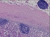
Fig. 1 (96k)
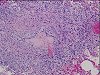
Fig. 2 (106k)
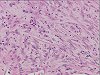
Fig. 3 (82k)
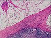
Fig. 4 (101k)
|
|
Abstract
SPONTANEOUS MULTICENTRIC RETROPERITONEAL FIBROMATOSIS IN A CYNOMOLGUS MONKEY (M. FASCICULARIS)
In a colony of cynomolgus monkeys a female animal not treated for several months developed signs of disease in form of recurrent diarrhea, anorexia, subcutaneous edema of the hindlegs and swelling of axillary lymph nodes. The animal was euthanised in moribund condition. In addition to the clinical observations necropsy revealed swelling of numerous body lymph nodes, marked thickening of the renal capsule and enlargement of spleen. Multiple grey-white nodules up to 10 mm in diameter and partly of firm consistency occurred in the thoracic and abdominal wall, mesentery, in all lobes of the lung and in the diaphragmatic and skeletal muscles. Histologic examination of selected tissue samples of the kidneys, lung, spleen, lymph nodes and of the nodules in the mesentery as well as in the musculature showed retroperitoneal fibromatosis with two morphologic patterns:
1. multicentric aggressively proliferating fibrous tissue with partly whorled or interwoven spindle shaped cells and 2. a sclerotic pattern with sparsely scattered mature fibroblasts within a densely packed bundle of collagen fibres. Local invasion of fibromatous tissue into the mesenchymal tissue of the musculature and into the perivascular tissue of the lung as well as into lymph node tissue was very prominent. Simian retroperitoneal fibromatosis is a fibroproliferative lesion which has many morphological and epidemiological similarities to human Kaposi's sarcoma which is highly associated with an immunodeficiency syndrome caused by viral infection (HIV, Simian retrovirus 2, SV40 and recently human herpes virus 8). In the case described here, virus isolation was not performed, but evidence of immunosuppression is given by the formation of multiple nodular infiltrates of mature lymphocytes with signs of germinal centre formation in various tissues and organs, especially prominent in the renal cortex. In addition, lymphocytic depletion and B-cell hyperplasia in lymph nodes and spleen were also observed and are as well considered to be indicative of immunosuppression. In animals without any signs of immunosuppression retroperitoneal fibromatosis has not been observed so far.
|
case index | << previous case | next case >>
|
|