
European Society of Toxicologic Pathology
GTP meeting 2000: Case No 2
case index | << previous case | next case >>
|
|



Abstract
Aim of the study
In this case report we describe the appearance of sarcocystosis in an SIV-infected rhesus macaque (Macaca mulatta).
Materials & methods
The animal was euthanized 25 weeks after SIV infection. Representative tissues were fixed in formalin, embedded in paraffin and stained with H&E.
Results
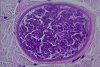
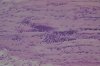
Apart from severe alterations in various organs due to opportunistic infections, the skeletal muscles appeared to be normal. In histology, the skin, skeletal muscles and the diaphragm were shown to contain numerous protozoic tissue cysts within remaining muscle fibres, with the fibres adjacent to the cyst's surface. The cysts were lined by a thick eosinophilic wall with long, regularly arranged, perpendicular cross striations. They contained large numbers of basophilic banana-shaped metrocytes and/or bradyzoites. The interstitial inflammatory reaction was mild and focal host reaction around parasitic cysts together with invading inflammatory cells could be seen. Multinucleated muscle cells indicating muscle regeneration were located next to the tissue cysts and inflammatory infiltrate, which consisted of lymphohistiocytic cells and mast cells accompanied by eosinophilic granulocytes.
Conclusions
With regard to the structure of the parasitic cysts, the shape of the metrocytes and the morphology of the cyst wall, we suspect Sarcocystis kortei to be the etiologic agent. Since infection with Sarcocystis spp. without contact to an intermediate host is not described in rhesus monkeys, the infection of this animal must have taken place before the onset of experiment. The number of sarcocysts and the amount of inflammatory response are of interest in this case, but the morphology of the cyst walls indicates maturity. It has to be considered that the inflammatory reaction is due to rupture of tissue cysts and the progressive failure of the immune system.