 |
 | |
| Species: |
Monkey |
| Strain/breeder: |
Callitrix jacchus (Marmoset) |
| Sex: |
Male |
| Age: |
3 years |
| Study type: |
Subchronic toxicity |
| Treatment: |
- |
| Animal status: |
Scheduled death, end of the study recovery period |
| Clinical findings: |
- |
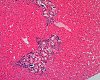
| Organ: |
Liver |
Macroscopic
finding(s): |
- |
| Staining: |
H&E |
| Literature: |
 |
Jones TC, et al. (1997) Veterinary pathology, 6th edition, p 641 |
|
Orikel TC, et al. (1972) Pathology of Simian Primates, Part II, p 76-103 |
|
Mehlhorn H, et al. (1993) Diagnose und Therapie der Parasitosen von Haus-, Nutz- und Heimtieren, 2. Aufl., pp 286-288 |
|
Thienpont D, et al. (1979) Diagnose von Helminthen, p 147 |
|
|
|
(98k)
|
|
Abstract
INFECTION WITH HEPATICOLA HEPATICA (CAPILLARIA HEPATICA) IN THE MARMOSET (CALLITHRIX JACCHUS)
Hepaticola hepatica (Capillaria hepatica) a nematode parasite has been reported from a wide variety of animals including primates, but this is the first case of an infection in a marmoset monkey. The histopathological findings of lesions produced by Hepaticola hepatica (Capillaria hepatica) in the liver of a 3-year old marmoset monkey will be presented.
The animal was in a 4-week subchronic toxicity study and the examination of the internal organs at the terminal necropsy was entirely unremarkable.
Microscopical examination of liver sections displayed areas of multiple ova, having caused tissue destruction. Subsequently, the lesions were invaded by connective tissue, monocytes and, in some cases, giant cells. The lesions were gradually replaced by scar tissue and were often accompanied by a mineralized center.
Other parasitic lesions caused by nematodes in Cynomolgus monkeys will be presented.
case index | << previous case | next case >>
|
|