
European Society of Toxicologic Pathology
ESTP Congress 2003: Case No 5
case index | << previous case | next case >>
|
|


Abstract
C.I. Rühl-Fehlert1, X. Palazzi2, M. Dorgerloh3, and C. Bomke4
1 Bayer HealthCare AG, Department of Toxicologic Pathology, Wuppertal, Germany
2 Bayer CropScience AG, Department of Pathology, Sophia Antipolis, France
3 Bayer CropScience AG, Institute for Ecotoxicology, Monheim, Germany
4 Dr. Knoell Consult, Mannheim, Germany
Key words: Fish, gonad, intersex
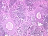
In a clinically healthy untreated adult fathead minnow of male external appearance (dark color, nuptial tubercles and dorsal fat pad, no ovipositor), ovaries were recorded at necropsy. Histopathology revealed in both gonads a mixture of testicular and ovarian tissue without clear separation of the two components. The testicular tissue consisted of seminiferous tubules containing spermatozoa and lined by spermatogenic cysts.
The ovarian tissue consisted of maturing follicles of various stages including late vitellogenic. The gonads had an oviduct which contained a huge amount of degenerated egg material/yolk plug. In addition, in the ovarian tissue a granulomatous inflammation due to an oocytic microsporidian infestation was observed.
In fish, sexual determination is particularly labile during a "critical phase of gonadal development" and also thereafter because bipotential germ cells may persist in mature gonads (Nakamura et al.: J Exp Zool 1998; 281: 362-372) which applies also to the gonochoristic species as the fathead minnow. Sex determination of fish can be influenced by various external stimuli as temperature, water pH, or social factors. In the present case it was not possible to determine the cause of this abnormality.