
European Society of Toxicologic Pathology
ESTP/GTP meeting 2002: Case No 9
case index | << previous case | next case >>
|
|

Abstract
W. KAUFMANN
BASF AG, Department of Product Safety, Regulations, Toxicology and Ecology, GV/T, Z 470, 67056 Ludwigshafen, Germany
Key words: zebra fish, parasites, myxozoa, myositis
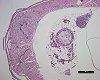
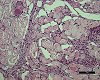
Adult zebra fish (9 months old) used in a fish full life cycle test (FFLC), have exhibited parasitic oocystes (up to a severe accumulation of different stages) in skeletal muscle fibre bundles and a reactive, granulomatous myositis. The protozoa are diagnosed as myxozoa - species which may primarily infest the central nervous system and are able to form xenomas (also: xenom or Xenoma tumour: a symbiotic complex formed by hypertrophying host cells and multiplying intra-cellular parasites, e.g. certain micro-sporidians). Xenomas in the spinal cord were not present in the two cases of infected fish investigated. However, xenoma-like lesions were found in the spinal root ganglia region with inflammatory response elements projecting into the surrounding somatic muscles. This xenoma development also involves the host immune response: a large number of macrophages gathered around the xenoma and infiltrate the infected cell, cross its plasma membrane, and destroys the spore mass. This phagocytic action leads to the formation of granulomas (J. L. MATTHEWS et al. (2001): J. EUKARYOT, Microbiol. 48 (2), pp 227-233).
In zebra fish, microsporidia infections may cause increased morbidity and mortality in fish which often appear clinically emaciated and lethargic.