
European Society of Toxicologic Pathology
ESTP/GTP meeting 2002: Case No 4
case index | << previous case | next case >>
|
|

Abstract
T. BLEIER and M. REINACHER
Department of Veterinary Pathology, University of Giessen, Frankfurter Str. 96, 35392 Giessen, Germany
Key words: feline, fibrosarcoma, vaccination, giant cells
A spayed female European short hair cat (9 years) developed an expanding mass over a period of 6 month at the shoulder. The cat was vaccinated at this location one year before the tumour developed. Further data about previous vaccinations are unknown. The tumour was surgically removed.
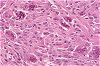
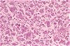
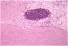
Histologically a fibrosarcoma was diagnosed. The tumour consisted of pleomorphic cells, multinucleated giant cells, a large area of necrosis, and clusters of lymphocytes at the tumour periphery. Tumour cells exhibited a high mitotic rate of 4 per high power field.
Four weeks later the cat was euthanised because of tumour recurrency. Together with the vaccination history, the histologic findings lead to the diagnosis of a vaccine-associated feline fibrosarcoma (COUTO et al. (2002), Vet Pathol 39: 33-41).