
European Society of Toxicologic Pathology
GTP meeting 2000: Case No 14
case index | << previous case | next case >>
|
|



Abstract
Aim of the study
An untreated male p53+/- mouse (TSG-p53 heterozygote, Taconic Farms, Inc., Germantown, NY, USA) from a life-span study was killed in a moribund condition at the age of 68 weeks. At necropsy, a firm nodule, measuring 0.12 cm in diameter, was detected within the duodenum and adjacent mesentery.
Materials & methods
All organs and macroscopic detectable lesions of the animal were trimmed, embedded in paraffin, and stained with H&E. A panel of immunohistochemial markers, a Masson Goldner-Trichrome stain and a Bielschowky-Novottny silver impregnation for reticulin were applied to several sections of the duodenal nodule.
Results
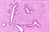
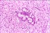
Microscopic examination of the duodenal nodule revealed local infiltration into the mesentery. The nodule contained glandular structures as well as a prominent sarcomatous component. Whereas the epithelial cells consisted of well differentiated goblet cells surrounding small lumens as well as low to tall columnar enterocytes with microvilli tips, the sarcomatous areas showed spindle shaped and round cells with light oval nuclei as well as anaplastic cells with giant nuclei. The Masson Goldner-Trichrome stain revealed a small amount of collagen fibres, and the reticulin stain showed a fine network of reticulin fibres. The immunohistochemical markers were positive for desmin, actin, and collagen type IV. Pan-cytokeratin (cko1, 5, 6, 8) was only positve in the goblet cell areas. There were negative reactions to ck7, F4/80, CD45R/B220, CD3, S-100, Factor VIII and Neurofilament 200.
Conclusions
Because of the histological cell type and the immunoreactivity pattern, the diagnosis of a poorly differentiated leiomyosarcoma with goblet cell metaplasia and reactive duodenal hyperplasia is proposed.