
European Society of Toxicologic Pathology
GTP meeting 2000: Case No 15
case index | << previous case | next case >>
|
|




Abstract
Aim of the study
In a long-term study using 2554 mice, the possible influence of parental radiation exposure on tumour development in the descendants was investigated. Female mice (C57/BL) were treated once by X-radiation two weeks prior to mating with untreated males (C3H), while the F1 descendants (B6C3F1) were either left untreated to observe the con-sequences of the preconceptual radiation exposure per se, or exposed to cyclosporine A. To guarantee clear and distinctive identification and to secure the individual parental origin of each mouse in the F1 generation, individually coded transponders (microchips) were implanted (s.c.) in all animals.
Materials & methods
The battery-free implantable micro-identification device (2 x 12 mm) contains an encoded microchip and a spool in a cylindrical glass capsule coated with a polypropylene cap on one side. Prepacked in the lumen of a sterile needle, they were injected subcutaneously in the dorsolateral back of the mice.
Results
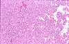
In single animals of this ongoing study, circumscribed subcutaneous nodules occurred at the site of implanted microchips. A firm, pale white nodule, up to 30 mm in diameter, completely embedding the microchip completely was found in a 39-weeks-old female C57BL mouse. Revealing a mixed histological appearance, the mesenchymal origin of the tumour cells was suspected by routine H&E staining and further immunohistochemical stainings are being performed to characterize this neoplasm.
Conclusions
Researchers/pathologists must be aware of foreign body tumorigenesis (microchip-induced neoplasms) possibly complicating the interpretation of data from carcinogenicity studies.