GTP meeting 2000: Case No 10
 | |
| Species: |
Rat |
| Strain/breeder: |
Wistar (Crl:WI(WU)BR) |
| Sex: |
Male |
| Age: |
122 weeks |
| Study type: |
Inhalation |
| Treatment: |
Clean air control |
| Animal status: |
Scheduled death, end of study |
| Clinical findings: |
Nodule, right ear |
| Organ: |
External ear |
Macroscopic
finding(s): |
Firm nodule within the right pinna, diameter: 0.8 cm |
| Staining: |
H&E |
| Literature: |
 |
Long PH, Leininger JR, Nold JB, Lieuallen WG (1993) Proliferative lesions of bone, cartilage, tooth, and synovium in rats. MST-2. Guides for Toxicologic Pathology. STP/ARP/AFIP, Washington, D.C. |
|
Yoshitomi K, Brown HR (1990) Ear and pinna. Pathology of the Fischer rat. Reference and atlas. Academic Press San Diego New York Boston, pp 227-238 |
|
|
|
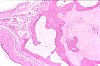
Fig. 1 (60k)
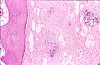
Fig. 2 (63k)
|
|
Abstract
CHONDRO-OSSEOUS METAPLASIA OF THE PINNA IN A WISTAR RAT
A male Wistar rat (Crl:WI(WU)BR) of the clean air control group from a long-term inhalation carcinogenicity study was terminally sacrificed at the age of 112 weeks. Clinical observations indicated a localized thickening of the right external ear. At necropsy, a firm nodule of the right pinna, measuring 0.8 cm in diameter, was recorded. Histopathological examination of decalcified tissue revealed that the nodule was composed of an outer rim of cartilage and mature lamellar bone with a core of fibrocartilage. The outer preexisting cartilage was narrow and irregular and was continuous on its inner aspect with mature lamellar bone enclosing marrow spaces with both haematopoietic cells and fatty tissue. The core of fibrocartilage was mainly composed of collagen fibres with interspersed chondrocytes. Parts of the fibrocartilage which exhibited a strong immunohistochemical staining for S-100 protein showed signs of myxomatous degeneration. Morphogenetically, the lesion is considered to represent the end-stage of a sequence starting with degeneration and splitting of the auricular cartilage, proliferation of perichondrial fibrous tissue and metaplastic transformation of the fibroblasts. Etiologically, the lesion may be related to traumatic injury, e.g. resulting from ear tattooing or ear tagging procedures. Occasional development of osteosarcomas at this site has been reported in Wistar rats.
case index | << previous case | next case >>
