|
| Guess What! - ESTP Case 10 |
The case shows H&E stained pictures from the oral cavity of a female Wistar rat aged 782 days, which was euthanized at the termination of a 24-months carcinogenicity study. No gross observations were reported.
| Click on the images below for a larger view. |
|
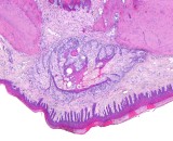
Fig. 1: H&E, x10
|
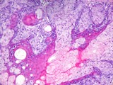
Fig. 2: H&E, x20
|
|
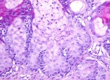
Fig. 3: H&E, x20
|
Morphologic Description
Unilaterally, in the region of right upper molar gingiva just beneath the mucosal surface is a well demarcated, non-encapsulated small area of well-differentiated lobular clustered sebaceous glands with acinar structure surrounded by a fine fibrovascular stroma. The sebaceous glands are unassociated with hair follicles and the glands have several small lobules consisting of acini composed of approximately 20 to 30 sebaceous cells. The sebaceous cells are young mature, degenerative and germinative cells. Most of the cells are young mature sebaceous cells, which are polyhedral in shape and each containing many small vacuoles within clear cytoplasm with a small, centrally-located nucleus. Single degenerative cells have swollen or ruptured cytoplasm filled with large vacuoles. At the base of the acini, there are cells with elongated, more basophilic cytoplasm interpreted as germinative cells. Additionally, randomly scattered but especially in the center of the gland are small islands of stratified squamous epithelium with a centrally located cavity.
Proposed Diagnosis
Unilateral Fordyce's granules of the upper molar gingiva in female Wistar rat
Differential Diagnoses and Discussion
Fourteen diagnoses were received, only four of them with the right diagnosis. Two of this senders diagnosed ectopic sebaceous glands, which is not wrong, but the sebaceous glands are most likely not of ectopic origin. Most of the senders diagnosed a sebaceous adenoma or carcinoma or at least a hyperplasia of the glands. To our knowledge, sebaceous adenomas or carcinomas are not yet described in the oral cavity of the rat and are also very rare entities in humans. There was a heavy discussion within the Guess What! Committee, if this lesion is still a normal Fordyce Granule or already a hyperplastic lesion. Because all participants of this committee did not see enough lesions of this character until now, the author decided to call it not hyperplastic, but still normal due to the facts, which can be taken from the literature. According to the descriptions in the published literature, there are more hints for normal than hyperplastic lesion, although a hyperplasia has to be considered as differential diagnosis.
So-called Fordyce's granules, which display morphologically normal sebaceous glands, are described in several species including humans and rats. They have to be differentiated from sebaceous gland adenomas and sebaceous gland hyperplasias, which are extremely rare entities in the oral cavity in humans and so far not yet described in rats or other species. The discussion is ongoing about the “ectopic” origin of Fordyce's granules, which are considered as ectopic sebaceous glands by some authors and as not ectopic sebaceous glands but normal oral mucosal adnexal glands by others, which is favored actually. Histologically, they are identical to their cutaneous counterpart but not associated with hair follicles.
In rats, there are, at present, only four reports of intraoral “ectopic” sebaceous glands in the literature (see references). In Holtzman and Long-Evans rats as well as in Wistar rats they were found in molar gingiva. Additionally, in F344 rats they were also detected in the gingiva of upper incisors. In F344 rats, the incidence of Fordyce's granules was markedly different when comparing sex, age, and site of lesion. They were very common in the midsagittal gingiva of the upper incisor in males (up to 56.3%) in contrast to females (up to 2.8%) and increased in incidence with age in both sexes. Though, in the molar gingiva they were very rare in both sexes and were found only in 1.2% of male and in 0.4% of female rats. In contrast, in male Wistar rats the incidence of Fordyce's granules in the upper molar gingiva was up to 12.5%. In addition, the most common site in rat molar gingiva is probably adjacent to the first molar. In most cases, Fordyce's granules were not grossly recognized but only histologically. Especially grossly recognized lesions may reveal a cystic dilation of ducts due to a sebum plug, which might be associated with an androgen-dependent increase of sebum production. Ultrastructurally, Fordyce's granules consist primarily of typical lipid-filled sebaceous cells. In addition to the incisor and molar also the buccal mucosa as well as the hard palate and tongue should be considered as possible sites of occurrence of Fordyce's granules in rats.
In humans, intraoral sebaceous glands on lips and buccal mucosa are an extremely common finding, which occur in both sexes with a higher incidence in men and in all age groups, although less often in young children.
References
- Bernick S, Bavetta LA (1962) The development of gingival sebaceous-like glands and cysts in rats of the Holtzman strain. Oral Surg Oral Med Oral Pathol 15:351-354
- Daley TD (1993) Intraoral sebaceous hyperplasia. Diagnostic criteria. Oral Surg Oral Med Oral Pathol 75:343-347
- Dreher A, Grevers G (1995) Fordyce spots. A little regarded finding in the area of lip pigmentation and mouth mucosa. Laryngorhinootologie 74:390-392
- Frandsen AM (1962) Sebaceous glands in the gingiva of the rat. Oral Biol 7:247-248
- Kaminagakura E, Andrade CR, Rangel AL, Coletta RD, Graner E, Almeida OP, Vargas PA (2003) Sebaceous adenoma of oral cavity: Report of case and comparative proliferation study with sebaceous gland hyperplasia and Fordyce's granules. Oral Dis 9:323-327
- Monteil RA (1981) Fordyce's spots: disease, heterotopia or adenoma? Histological and ultrastructural study. J Biol Buccale 9:109-128
- Rulli MA, Martinelli C (1971) Free sebaceous glands in the gingival of some rats. Archs Oral Biol 16:831-832
- Yoshitomi K, Brown HR, Eustis S (1990) Fordyce's granules of the incisor and molar gingiva in F344 rats. Vet Pathol 27:432-438
|
|