|
| Guess What! - ESTP Case 20 |
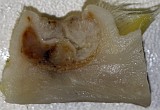
This male Wistar rat from the control group of a carcinogenicity study was killed at an age of 733 days. At necropsy, the animal showed at the right side of the thorax a 1.5 cm in diameter sized skin lesion.
| Click on the images below for a larger view. |
|
Fig. 1: Macroscopic findings
|
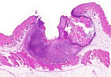
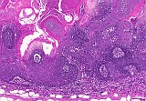
Fig. 2: H&E, x1
|
|
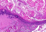
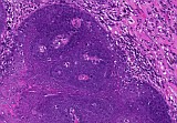
Fig. 3: H&E, x10
|
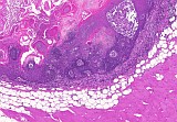
Fig. 4: H&E, x10
|
|
Fig. 5: H&E, x20
|
Fig. 6: H&E, x40
|
|
Fig. 7: H&E, x40
|
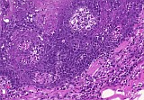
Fig. 8: H&E, x10
|
|
Fig. 9: H&E, x20
|
Fig. 10: H&E, x40
|
|
Fig. 11: H&E, x10
|
Fig. 12: H&E, x10
|
|
Fig. 13: H&E, x20
|
Fig. 14: H&E, x40
|
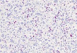
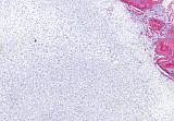
Morphologic Description
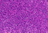
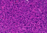
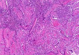
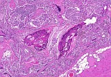
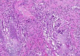
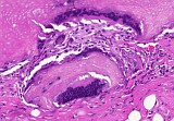
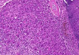
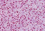
The dermis showed a well demarcated unencapsulated mass (Figs. 1&2) consisting of nodules of multilayered epithelial cells surrounding a central lumen filled with keratin and ghost cells (Figs. 3-5). Keratinization is abrupt. In some regions, trichogenic differentiation is recognizable. In some areas the basal cells exhibited atypia and a high mitotic rate (Figs. 6&7). Areas adjacent to the mass showed a mainly unimorphic cell population consisting of multinucleated giant cells (Figs. 8-10). These cells were positive for the macrophage marker ED1/CD68 (Fig. 16) but negative for the cell proliferation marker Ki67 (Fig. 17). Pan-cytokeratin (Ck 1, 5, 6, 8) positive staining was not present in this area (Fig. 18). Other areas showed free keratin with a granulomatous inflammation, multinucleated giant cells trying to engulf the keratin, mineralization, and osseous metaplasia (Figs. 11-14).
Proposed Diagnosis
Tumor, hair follicle, benign; pilomatricoma type
with focal atypia, free intradermal keratin, granulomatous inflammation, and numerous partly unimorphic multinucleated giant cells.
| Click on the images below for a larger view. |
|
Fig. 15: H&E, x20
|
Fig. 16: IHC_ED1_CD68, x20
|
|
Fig. 17: IHC_Ki67, x40
|
Fig. 18: IHC_PanCK_Mix, x10
|
Discussion
13 diagnoses were received for this case.
5 persons suggested a keratoacantoma (synonym infundibular keratinizing acanthoma) and three persons a tumor, hair follicle, benign; pilomatricoma type (synonym pilomatricoma and as human medical term: pilomatrixoma or epithelioma calcificans Malherbe). Furthermore, three persons suggested a malignant tumor (malignant trichoepithelioma, basal cell carcinoma, squamous carcinoma).
Though keratoacantomas and hair follicle tumors, pilomatricoma type could look quite similar, due to the evident ghost cells, we think this to be a hair follicle tumor. Furthermore, structures resembling hair follicles could be seen (Fig. 5). Within this tumor areas were visible which showed atypia and a high mitotic rate. However, no invasion of subcutis or desmoplasia was present. Furthermore, though described to occur in domestic animals, according to the recent INHAND nomenclature no malignant hair follicle tumors in rodents were described so far. These tumors might be diagnosed as basal cell or squamous carcinoma.
Basal cells carcinomas should show invasion into basement membrane and surrounding tissue with evidence of indistinct demarcation and heterogeneous cytological and growth pattern. They are traditionally considered epidermal neoplasms although it appears much more likely that most of them originate from hair germ epithelium, which is the reason why many of these tumors show some differentiation into sebaceous cells. Basal cell tumors should not show trichogenic differention. Basal cell carcinomas are usually of low grade malignancy and metastases are rare.
Squamous carcinomas are composed of islands or cords of cells that penetrate the basal lamina and invade the dermis. The tumor cells might reach the subcutis or subcutaneous muscle. There is some evidence of squamous differentiation with atypia and frequent mitotic figurs, although its extent is variable.
Areas adjacent to the tumor showed a mainly unimorphic cell population consisting of multinucleated giant cells (Figs. 8-10). Presumably this led to the diagnosis of a rhabdomyosarcoma as suggested by one person. Rhabdomyosarcomas are tumors frequently showing necrosis and hemorrhage. They are highly pleomorphic with rhabdomyoblasts, immature spindle and strap-like cells, mononucleated, rounded and polygonal cells. Rhabdomyoblasts are characterized by eosinophilic fibrillar cytoplasm with myofilaments, glycogen inclusions, cross striations, immunocytochemical staining for myoglobin and ultrastructural evidence of Z bands. They show a high mitotic activity with abnormal mitotic figures. They are locally infiltrative with frequent distant metastases. Since the multinucleated cells in this tumor were positive for the macrophage marker ED1/CD68 we excluded this tumor.
The last person suggested a collision tumor with parts of all three germ cell layers, differentiation to squamous carcinoma (originating from hair follicle), giant cell sarcoma, osteosarcoma, and myosarcoma. Originally we were unsure about the unimorphic cell population consisting of multinucleated giant cells adjacent to the tumor. But immunohistochemistry helped to solve this problem. Since these cells were all positive for the macrophage marker ED1/CD68 and negative for the cell proliferation marker Ki67, we ruled out that these cells were tumor cells. Furthermore, additional trimming of remaining tumor tissue revealed that these cells were admixed with keratin remnants (Fig. 15). Therefore, we think that these cells are part of the granulomatous inflammation directed against the free keratin within the dermis.
References
- Greaves P, Chouinard L, Ernst H, Mecklenburg L, Pruimboom-Brees IM, Rinke M, Rittinghausen S, Thibault S, Von Erichsen J, Yoshida T (2013) Proliferative and non-proliferative lesions of the rat and mouse soft tissue, skeletal muscle and mesothelium. J Toxicol Pathol 26(3): 1S-26S
- Mecklenburg L, Kusewitt D, Kolly C, Treumann S, Adams ET, Diegel K, Yamate J, Kaufmann W, Müller S, Danilenko D, Bradley A (2013) Proliferative and non-proliferative lesions of the rat and mouse integument. J Toxicol Pathol 26 (3): 27S–57S
- Le Boit PE (ed.) (2006) World Health Organization Classification of Tumours. Pathology and Genetics of Skin Tumours. IARC Press, Lyon.
|
|