|
| Guess What! - ESTP Case 19 |
The control male CrlGlxBrl Han:WI (SPF) rat aged 36 weeks, was killed at the end of a 26-week systemic oral gavage toxicity study. At necropsy the animal showed a thickening of the ear, tagged with a metal clip.
| Click on the images below for a larger view. |
|
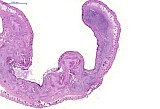
Fig. 1: H&E, x1
|
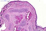
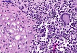
Fig. 2: H&E, x5
|
|
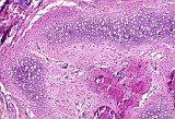
Fig. 3: H&E, x10
|
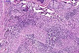
Fig. 4: H&E, x10
|
|
Fig. 5: H&E, x40
|
Morphologic Description
Ear, histologically, severe destruction of the normal cartilaginous plate, formation of new cartilaginous nodules and osseous metaplasia with a severe multifocal granulomatous inflammatory reaction causing extensive thickening (Fig.1) of the ear. The granulomatous inflammation (Fig 4) is characterized by a central core of degenerated and viable neutrophils surrounded by epitheloied macrophages, lymphocytes and plasma cells, fibroblasts and fibrocytes and occasionally multinuclear giant cells (Fig. 5). Multifocal proliferating cartilaginous nodules containing large swollen chondrocytes with round to oval nuclei (Fig 2, 3 and 5) are separated by fibrous tissue containing few lymphocytes and plasma cells. Occasional bone formation (osseous metaplasia) and mineralization are present (Fig 2 and 3).
Proposed Diagnosis
Ear, Auricular chondritis (auricular chondropathy) Multifocal granulomatous inflammation with destruction of the cartilaginous plate accompanied by formation of cartilaginous nodules and osseous metaplasia.
Discussion
16 diagnoses were received for this case. By far the most contributors suggested auricular chondritis (auricular chondrophathy) (5), granulomatous inflammation (4) with secondary cartilage proliferation and bone formation (1 contributor) and with giant cells and dystrophic calcification (another contributor), or chondroid degeneration, granulomatous inflammation and osseous metaplasia and fibroplasia (1).
Other contributors suggested Expression of 'arthritis' in the ear with secondary reactive changes, such as bone formation (1), Chondrodysplasia with lymphoplasmacytic inflammation intermingled with Langhans Giant cells (1), metaplastic mineralization accompanied by chronic histiocytic inflammation characterized by multifocal giant cell formation (1), othematoma with sequestered cartilage and chronic inflammation (1) and granulomatous dermatitis (2).
Malignant fibrous histiocytoma was suggested as differential diagnosis (1). Foreign body reaction e.g following collagenolysis (1), Myobacterium spp (1), trauma (1) and autoimmunity (1) were suggested as possible etiologies.
Spontaneous auricular chondiritis (auricular chondropathy) is characterized grossly by unilateral or bilateral nodular to diffuse thickening of auricles in aged rat. Histopathologically, the lesion has been described as a multifocal granulomatous inflammation with destruction of the cartilaginous plate accompanied by formation of cartilaginous nodules and osseous metaplasia. The disease has been observed in several rat strains, including S-D and Wistar rats and C57BL/6 mice (McEwen and Barsoum 1990; Meingassner 1991; Kitagaki et al., 2002; Kitagaki and Hirota 2007). Trauma and infectious agents have been considered as possible underlying causes, but it may be an immunologically mediated disease (Percy and Barthold, 2007).
The lesion develops initially in the tagged ear long after that the traumatic insults form the ear tagging procedure is healed and then quite often becomes bilateral affecting also the non-tagged ear. Laceration/loss of metal ear tags heals without ear deformity and nylon ear tags, ear punching/notching and color markings on the dorsum, and traumatic insults to the pinnal cartilage does not cause similar lesions (McEwen and Barsoum 1990; Meingassner 1991; Kitagaki et al., 2002; Kitagaki and Hirota 2007). Meingassner, 1991 suggested that cartilage matrix components are involved in an autoimmune process resulting in a granulomatous destructive auricular chondropathy since they found IgG and complement deposits in the matrix of the marginal areas of new forming cartilaginous nodules and at the destruction sites of cartilage. Kitagaki and Hirota, 2007 investigated the pathogenesis of auricular chondritis caused by metal ear tagging in C57BL/6 mice based on the hypothesis that it is caused by metal ions release from the metal tag. They found that increased concentrations of copper and iron ions in the tagged ear was accompanied by up-regulation of metallothionein- I and -II mRNA, infiltration of CD4T lymphocytes, macrophages, neutrophils and mast cells and expression of Th1 cytokines such as IFN-γ, TNF-α, and IL-2. The resulting autoimmune response may than lead to the progression of auricular chondritis as an autoimmune disease.
Walkers et al., 1987 observed an increase of malignant tumors predominantly osteosarcomas at the site of the metal ear tags in a 2-yr carcinogenesis study in male Wistar rats. Elementary analysis of the tags showed that they were mainly composed of nickel and copper. Persistent inflammatory tissue reaction to the metallic device was considered to be an important factor in tumor development.
In humans auricular chondritis after high ear piercing have been described to be associated with poor training of the piercer, use of benzalkonium chloride as a preparation agent and infection with Pseudomonas spp. (More et al., 1999). After removal of the piercing, adequate wound treatment and systemic antibiotics the lesions commonly healed without deformations. Although laboratory rats are susceptible to experimental infections with Mycobacterium spp., naturally occurring infections are very rare and lesions affecting the cartilaginous plate in the ear, subcutaneous tissue and/or skin in the rat have not been reported in the literature.
With expression of 'arthritis in the ear' the contributor likely referred to the inflammatory destruction of elastic ear cartilage with multifocal nodular chondritis that develops on occasion, in Sprague-Dawley (S-D) rats after the onset of type II collagen-induced arthritis (McCune et al., 1982). In this case the ear lesions were not induced by type II collagen, which is the major collagen type in cartilage, injections.
Chondrodysplasia is a general term for a disorder of the development if cartilage. Severe, progressive growth plate chondrodysplasia has been described as an accompanying lesion in the tl rat which has a frame shift mutation in the Csf-1 gene that renders it null, resulting in severe osteopetrosis (Devraj et al 2004). The lesions in the growth plate were not accompanied by overt inflammation and an involvement of the auricular cartilaginous plate was not mentioned in the literature.
The terminology metaplastic mineralization is not a common established term in medicine and/or toxicopathology. Metaplasia is the reversible replacement of one differentiated cell type with another mature differentiated cell type. Whereas mineralization can be divided into dystrophic calcification/mineralization which occurs despite normal serum calcium levels in dying tissue and metastatic calcification/mineralization which occurs in normal tissue due to hypercalcemia (Kumar et al., 2010).
Othematoma – is described as a cystic swelling of the pinna of the ear due to hemorrhage without serious inflammation and is a routine finding in dog and cats and may be caused by traceable traumas or micro-traumas (Matousek JL, 2004). According to the literature othematoma is not a common described finding in the rat.
Since the granulomatous inflammation is located in the cartilaginous plate of the ear and not the skin, the term dermatitis is considered inapplicable.
Malignant fibrous histiocytoma (FIBROSARCOMA, PLEOMORPHIC, goRENI) is a solid fibrous mass with variable histology ranging from storiform pattern of uniform plump spindle cells, small rounded cells, to a highly pleomorphic pattern of bizarre spindle cells with variable mitotic activity and tumor giant cells. Abundant but variable amount of interstitial collagen which may show myxomatous zones is present. Formation of cartilaginous nodules, osseous metaplasia and overt granulomatous inflammation which are a characteristic feature in this case are not a diagnostic feature of malignant fibrous histiocytoma. The tumor shows widespread infiltration and invasion of local tissues. Subcutaneous implantation of inert plastics, metals and other materials of certain dimensions can likewise give rise to sarcomas around implantation sites in rodents, the so called 'Oppenheimer effect' or 'solid state' carcinogenesis [goRENI (Oppenheimer et al., 1953; Autian 1973; Brand et al., 1976; Kirkpatrick et al., 2000; Hahn et al., 2002)].
References
- Aharinejad S1, Grossschmidt K, Franz P, Streicher J, Nourani F, MacKay CA, Firbas W, Plenk H Jr, Marks SC Jr. (1999) Auditory ossicle abnormalities and hearing loss in the toothless (osteopetrotic) mutation in the rat and their improvement after treatment with colony-stimulating factor-1. J Bone Miner Res 14: 415-23
- Autian J (1973) The new field of plastics toxicology--methods and results. CRC Crit Rev Toxicol 2: 1–40
- Brand KG, Johnson KH, Buoen LC (1976) Foreign body tumorigenesis. CRC Crit Rev Toxicol 4: 353–394
- Hahn FF, Guilmette RA, Hoover MD (2002) Implanted depleted uranium fragments cause soft tissue sarcomas in the muscles of rats. Environ Health Perspect 110: 51–59
- Devraj K1, Bonassar LJ, MacKay CA, Mason-Savas A, Gartland A, Odgren PR. (2004) A new histomorphometric method to assess growth plate chondrodysplasia and its application to the toothless (tl, Csf1(null)) osteopetrotic rat. Connect Tissue Res 45:1-10
- goRENI; Fraunhofer ITEM, Hannover, Germany
- Kirkpatrick CJ, Alves A, Köhler H, Kriegsmann J, Bittinger F, Otto M, Williams DF, Eloy R (2000) Biomaterial-induced sarcoma: A novel model to study preneoplastic change. Am J Pathol 156: 1455–1467
- Oppenheimer BS, Oppenheimer ET, Stout AP (1953) Carcinogenic effect of imbedding various plastic films in rats and mice. Surg Forum 4: 672–676
- Kitagaki M, Hirota M. (2007) Auricular chondritis caused by metal ear tagging in C57BL/6 mice. Vet Pathol 44: 458-66.
- Kitagaki M, Suwa T, Yanagi M, Shiratori K. (2003) Auricular chondritis in young ear-tagged Crj:CD(SD)IGS rats. Lab Anim 37: 249-53
- Kopera D1, Soyer HP, Smolle J, Kerl H. (2000) "Pseudocyst of the auricle", othematoma and otoseroma: three faces of the same coin? Eur J Dermatol 10: 451-4
- Kummar, Abbas, Fausto, Aster. (2010) Robbins and Cotran: Pathologic Basis of Disease. 8 Ed. Saunders Elsevier. pp 10, 38-39
- Matousek JL, (2004) Diseases of the ear pinna. Vet Clin North Am Small Anim Pract 34 (2): 511-540
- McCune WJ, Schiller AL, Dynesius-Trentham RA, Trentham DE. (1982) Type II collagen-induced auricular chondritis. Arthritis Rheum 25: 266-73
- McEwen BJ, Barsoum NJ. (1990) Auricular chondritis in Wistar rats. Lab Anim 24: 280-3
- Meingassner JG. (1991) Sympathetic auricular chondritis in rats: a model of autoimmune disease? Lab Anim 25: 68-78
- More DR, Seidel JS, Bryan PA. (1999) Ear-piercing techniques as a cause of auricular chondritis. Pediatr Emerg Care 15: 189-92
- Percy DH and Barthold SW (2007) Pathology of laboratory rodents and rabbits. 3rd Ed. Blackwell Publishing p.167
- Waalkes MP, Rehm S, Kasprzak KS, Issaq HJ. (1987) Inflammatory, proliferative, and neoplastic lesions at the site of metallic identification ear tags in Wistar [Crl:(WI)BR] rats. Cancer Res 47: 2445-50
|
|