|
| Guess What! - ESTP Case 8 |
The case is from a 10-week-old female Syrian golden hamster (Han:AURA). The macroscopic finding was: slight focal thickening of the skin, lateral abdominal region.
| Click on the images below for a larger view. |
|
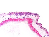
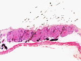
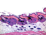
Fig. 1: H&E, x25
|
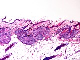
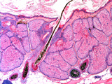
Fig. 2: H&E, x100
|
Morphologic Description
The present case is characterized by a focal dermal clustering of prominent sebaceous glands associated with large hair follicles which extend into the subcutaneous fat tissue. The coarse hairs are pigmented and in some areas, clumps of dermal pigment are bordering on the pilosebaceous units.
Proposed Diagnosis
(Female) flank organ
Synonyms: costovertebral scent gland, costovertebral pigmented spot, organ of Kupperman
Differential Diagnoses and Discussion
The majority of the contributors (9/21) diagnosed a (multi)focal sebaceous (gland) hyperplasia of the skin. This is the morphologically correct diagnosis if the change would have been located at a different site of the body. We have to admit that the macroscopic finding was somewhat misleading because it mentioned only a unilateral lesion.
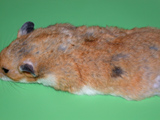
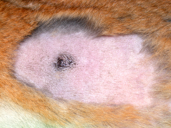
The flank organ was recognized by 7 contributors. The paired flank organ of the Syrian hamster can be seen macroscopically after the hair has been removed as grey (female) or black (male) spot (Figs. 3 and 4). At necropsy, it is also visible as greyish spot on the bottom side of the skin. In both male and female hamsters, the flank organ is not well developed until the onset of puberty. Thereafter, under the influence of sex hormones (androgens), the sexual dimorphism becomes evident resulting at an age of about 3 months in a volume of approximately 23 mm3 for the male and 8 mm3 for the female flank organ. The flank organs of adult males are about twice the size in diameter (5 to 10 mm) than of females and bulge slightly above the surrounding skin in comparison to the females (Fig. 5). Histologically, they are composed of giant sebaceous glands discharging into large hair follicles (Fig. 6). The follicles produce coarse terminal hair and especially in males are surrounded by a dense network of pigment-secreting melanocytes (Fig. 7). In females, pigmentation of the glandular area is much weaker than in males.
The flank organs are considered to be secondary sex organs. Males are reported to investigate female flank organs prior to mating and they mark their territory by a porphyrin-containing secretion of the glands. Flank organs also appear to play a role in the conversion of testosterone to dihydrotestosterone, indicating the importance of androgens for hair growth and flank organ growth. In young males with aspermatogenesis (Fig. 8a), the flank organ shows a reduced size and resembles the one of female hamsters (Fig. 8b).
The flank organs of the Syrian golden hamster have been widely used as a model for the human sebaceous gland by investigating effects of systemically or topically applied hormones, antiandrogens and retinoids. Assays using testosterone-stimulated female hamsters test the ability of antiandrogenic drugs to inhibit the initiation of androgen-dependent growth, whereas the reduction of established growth is examined with the male hamster assays. In these tests, the in vivo flank organ size is usually expressed as the product of the greatest longitudinal and greatest transverse diameter of the palpable organ. The sebaceous gland volumes of the flank organ can be calculated from computer-assisted planimetry of serially sectioned specimens or by applying modern in vivo ultrasonic imaging techniques. The relevance of the hamster flank organ model for man, however, has also been questioned since the sebaceous gland of the hamster flank organ is apparently more sensitive to antiandrogens than the human sebaceous gland.
Three of the 21 persons who submitted a diagnosis had seen or suspected a benign skin tumour, a trichoepithelioma, which could have been related to hamster polyoma (papova) virus (HaPV) infection. This tumour type, however, is mainly composed of concentrically arranged nests of epithelial cells around hairshaft material or optically empty spaces, while mature hair shafts and infundibula are missing. A significant sebaceous component as in the flank organ is also missing in trichoepitheliomas.
One contributor diagnosed a basal cell tumour. Such lesions are typically composed of a uniform population of small basophilic cells resembling basal cells of the epidermis. They form palisades, ribbons and anastomosing cords and also lack hair structures and a significant sebaceous component.
Another contributor submitted the diagnosis of a sebaceous adenoma. The sebaceous glands indeed are the main histological features of the flank organ. However, they are regularly associated with hair follicles and the normal skin architecture is preserved. By an expansile endophytic or exophytic growth pattern, sebaceous adenomas usually produce a marked disturbance of the normal skin structures.
With respect to tumorigenesis, the flank organ has been reported to be most resistant to tumour production by chemical carcinogens. This has been attributed to the large sebaceous glands in the flank organ which rid themselves and the hair follicles rapidly of carcinogens (flushing action).
| Click on the images below for a larger view. |
|
Fig. 3: Male Syrian golden hamster
|
Fig. 4: Male Syrian golden hamster
|
|
Fig. 5: H&E, x25
|
Fig. 6: H&E, x100
|
|
Fig. 7: H&E, x200
|
Fig. 8a: H&E, x200
|
|
Fig. 8b: H&E, x50
|
References
- Kupperman H (1944) Hormone control of a dimorphic pigmentation area in the golden hamsters (Cricetus auratus). Anat Rec 88: 442
- Ghadially FN, Ghadially R (1996) Tumours of the skin. In: Turusov VS, Mohr U (eds) Pathology of tumours in laboratory animals. Vol. III – Tumours of the hamster. International Agency for Research on Cancer, Lyon, France
- Lucky AW, Eisenfeld AJ, Visintin AA (1985) Autoradiographic localisation of tritiated dihydrotestosterone in the flank organ of the albino hamster. J Invest Dermatol 84: 122-125
- Algard FT, Dodge AH, Kirkman H (1966) Development of the flank organ (scent gland) of the Syrian hamster. II. Postnatal development. Am J Anat 118: 317-326
- Combettes C, Durand-Seme V, Querleux B, Saint-Leger D, Leveque JL (1989) Imaging the hamster flank organ by an ultrasonic technique: a new approach to animal tests. Br J Dermatol 121: 689-699
- Franz TJ, Lehman PA, Pochi P, Odland GF, Olerud J (1989) The hamster flank organ model: is it relevant to man? J Invest Dermatol 93: 475-479
- Weissmann A, Bowden J, Frank B, Horwitz SN, Frost P (1984) Morphometric studies of the hamster flank organ: an improved model to evaluate pharmacologic effects on sebaceous glands. J Invest Dermatol 82: 522-525
- Percy DH, Barthold SW (2001) Pathology of Laboratory Rodents & Rabbits. Iowa State University Press, 2nd edition, page 168
|
|