 |
 | |
| Species: |
Rat |
| Strain/breeder: |
Wistar (Janvier, 53940 Le Genest St. Isle, France) |
| Sex: |
Male |
| Age: |
Approximately 20 weeks |
| Study type: |
Modified Segment I study |
| Treatment: |
13 Weeks oral (gavage) |
| Animal status: |
Scheduled sacrifice, termination of study |
| Clinical findings: |
None |
| Organ(s): |
Testis (3 cut levels) |
Macroscopic
finding(s): |
None |
| Staining: |
H&E |
| Special request: |
Toxicological assessment of findings |
|
|
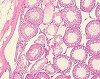
Fig. 1 (106k)
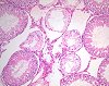
Fig. 2 (88k)
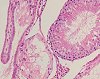
Fig. 3 (78k)
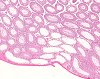
Fig. 4 (98k)
|
|
Abstract
Segmental (focal) tubular atrophy in rat testes (induced lesion)
S. HALM and C. GESCHWILL
Abbott GmbH & Co. KG, Toxicology, Knollstrasse 50, 67008 Ludwigshafen, Germany
Key words: testicular changes, tubular atrophy, rat
The testes of Wistar rats of a segment I study (14-week treatment) were cut into 5 levels with a template, from the dorsal part of the testis to the ventral part. All 5 levels were embedded and the paraffin sections were prepared and stained with H&E. For quantification, all slides were examined for altered tubules which were counted in both testes. Altered tubules were defined as containing more than one vacuole per tubule, with changes ranging from decrease in height of the germinal epithelium to tubular atrophy and/or tubules with calcified sperm, summarized as tubular atrophy. The total number of sectioned tubules in all 5 levels was determined in several testes. The results were compiled as the number of altered (sectioned) tubules (per animal and testis) and as the percentage of altered tubules in relation to total tubules per testis.
The testis revealed multifocal (segmental) tubular atrophy in association with dilation of the rete testis. The number of altered tubules in the treated animals was low in relation to the total number of sectioned tubules. In most testes, the percentage of segmental (focal) tubular atrophy was less than 10% even in animals with massive tubular atrophy. The majority displayed only minimal to slight tubular atrophy with less than 1% of the tubules altered. Thus, testicular alteration of multifocal or segmental tubular atrophy was generally slight and did not have any impact on fertility.
case index | << previous case | next case >>
|
|