 |
 | |
| Species: |
Mouse |
| Strain/breeder: |
B6C3F1 mouse, Charles River |
| Sex: |
Male |
| Age: |
18 months |
| Study type: |
Carcinogenicity study |
| Treatment: |
Control |
| Animal status: |
Survivor |
| Clinical findings: |
|
| Organ(s): |
Liver |
Macroscopic
finding(s): |
Mass, left medial lobe, cystic,
10 mm x 5 mm, yellow-white |
| Staining: |
H&E |
|
|
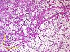
Fig. 1 (124k)
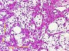
Fig. 2 (119k)
|
|
Abstract
Ito cell tumour in the liver of a B6C3F1 mouse
K. KÜTTLER
BASF Aktiengesellschaft, Product Safety, Regulations, Toxicology and Ecology, GV/T, Z470, 67056 Ludwigshafen/Rhein, Germany
Key words: Ito cell tumour, mouse, liver
At terminal sacrifice, an untreated male B6C3F1 mouse from a 18 month-carcinogenicity study showed a 10 x 5 mm yellow-white mass in the left medial liver lobe. Microscopically, the mass consisted mainly of vacuolated or signet-ring-shaped cells. In the peripheral region of the mass small spindle-shaped cells proliferated between normal liver cell plates and the vacuolated cells. Remnants of atrophic liver cell plates and isolated hepatocytes were distributed among the tumour cells. Collagen fibres were seen especially around the spindle-shaped cells. Immunohistochemically, the tumour cells stained positive for desmin, alpha-smooth muscle actin, laminin and tenascin. The ultrastructural characteristics of the tumour cells were consistent with Ito cells. Therefore, the tumour was diagnosed as an Ito cell tumour. Ito cell tumours are rarely seen in the liver. Evaluation of historical data from BASF over the last 20 years showed two further cases in the mouse and one in the liver of a Wistar rat.
|
case index | << previous case | next case >>
|
|